Details of the Target
General Information of Target
| Target ID | LDTP05802 | |||||
|---|---|---|---|---|---|---|
| Target Name | Serine/arginine-rich splicing factor 6 (SRSF6) | |||||
| Gene Name | SRSF6 | |||||
| Gene ID | 6431 | |||||
| Synonyms |
SFRS6; SRP55; Serine/arginine-rich splicing factor 6; Pre-mRNA-splicing factor SRP55; Splicing factor, arginine/serine-rich 6 |
|||||
| 3D Structure | ||||||
| Sequence |
MPRVYIGRLSYNVREKDIQRFFSGYGRLLEVDLKNGYGFVEFEDSRDADDAVYELNGKEL
CGERVIVEHARGPRRDRDGYSYGSRSGGGGYSSRRTSGRDKYGPPVRTEYRLIVENLSSR CSWQDLKDFMRQAGEVTYADAHKERTNEGVIEFRSYSDMKRALDKLDGTEINGRNIRLIE DKPRTSHRRSYSGSRSRSRSRRRSRSRSRRSSRSRSRSISKSRSRSRSRSKGRSRSRSKG RKSRSKSKSKPKSDRGSHSHSRSRSKDEYEKSRSRSRSRSPKENGKGDIKSKSRSRSQSR SNSPLPVPPSKARSVSPPPKRATSRSRSRSRSKSRSRSRSSSRD |
|||||
| Target Bioclass |
Other
|
|||||
| Family |
Splicing factor SR family
|
|||||
| Subcellular location |
Nucleus
|
|||||
| Function |
Plays a role in constitutive splicing and modulates the selection of alternative splice sites. Plays a role in the alternative splicing of MAPT/Tau exon 10. Binds to alternative exons of TNC pre-mRNA and promotes the expression of alternatively spliced TNC. Plays a role in wound healing and in the regulation of keratinocyte differentiation and proliferation via its role in alternative splicing.
|
|||||
| Uniprot ID | ||||||
| Ensemble ID | ||||||
| HGNC ID | ||||||
| ChEMBL ID | ||||||
Target Site Mutations in Different Cell Lines
Probe(s) Labeling This Target
ABPP Probe
| Probe name | Structure | Binding Site(Ratio) | Interaction ID | Ref | |
|---|---|---|---|---|---|
|
P1 Probe Info |
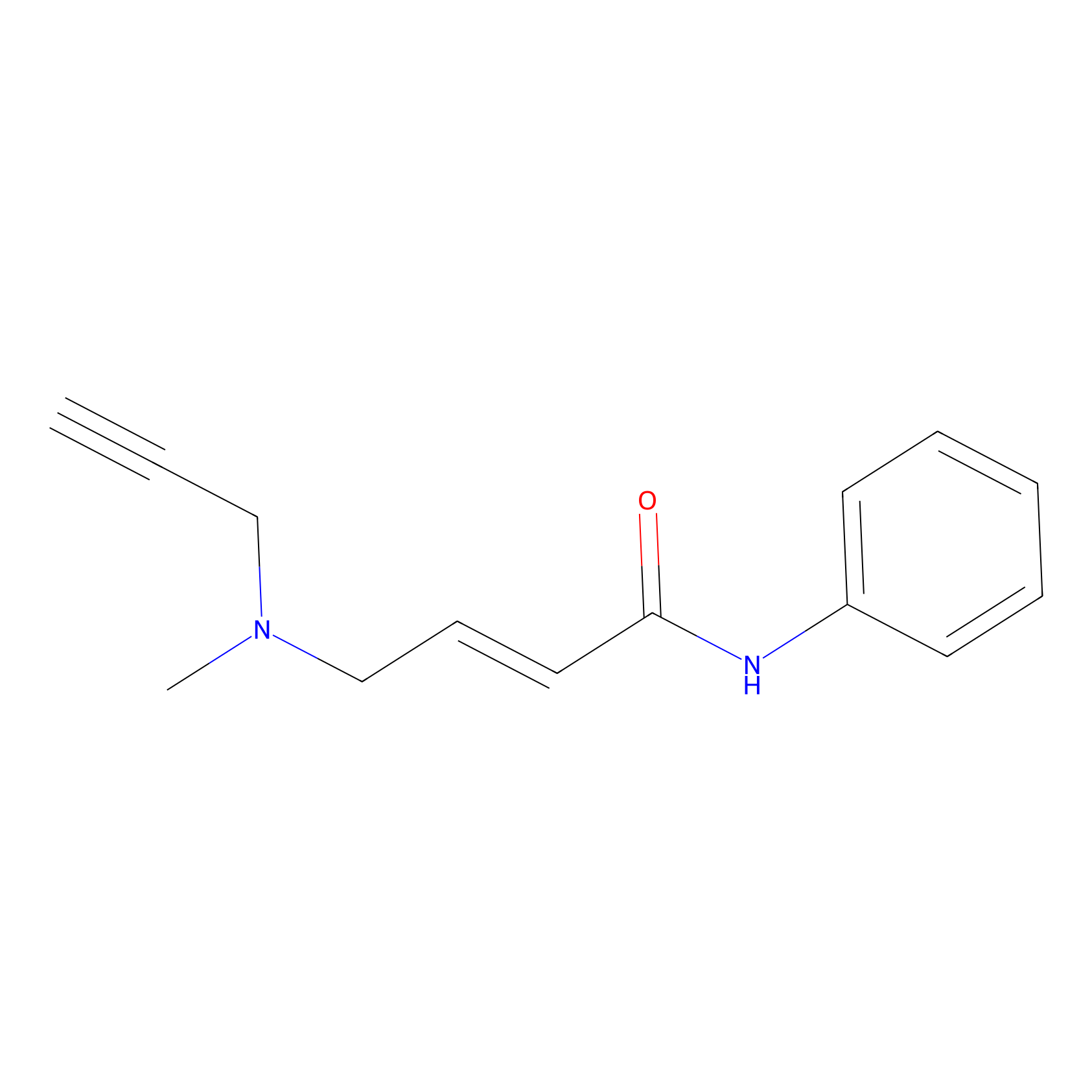 |
1.70 | LDD0448 | [1] | |
|
P3 Probe Info |
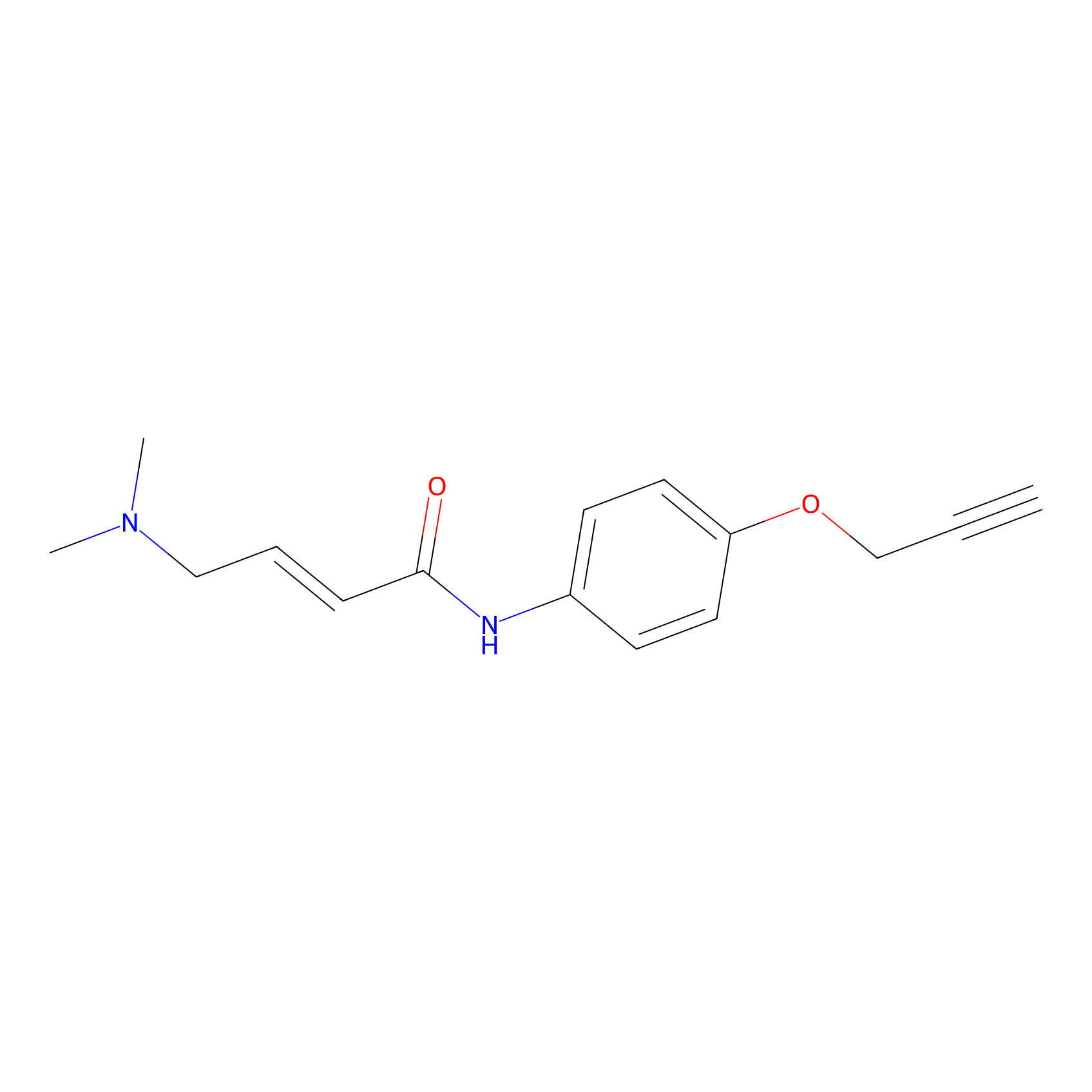 |
1.94 | LDD0450 | [1] | |
|
C-Sul Probe Info |
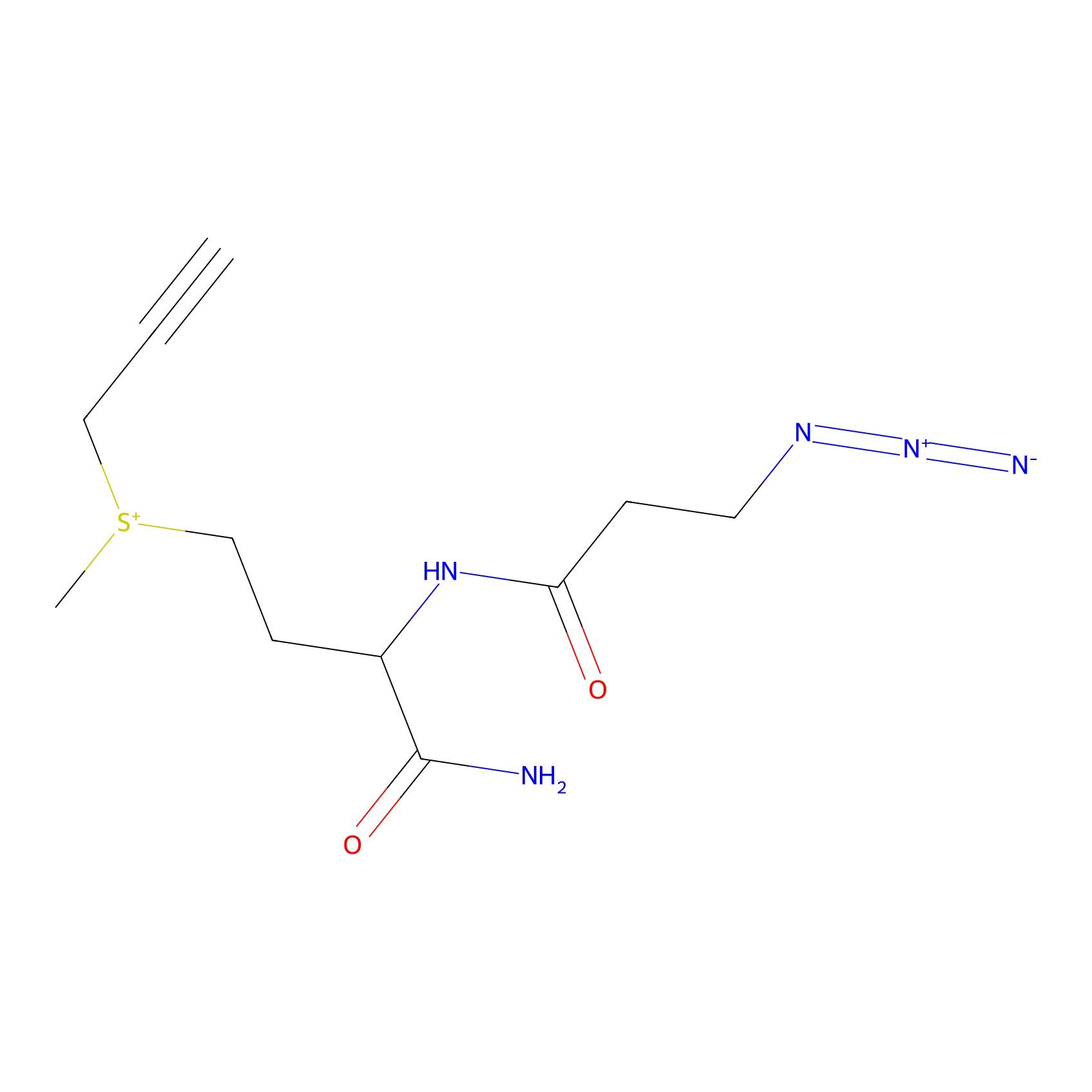 |
7.24 | LDD0066 | [2] | |
|
YN-1 Probe Info |
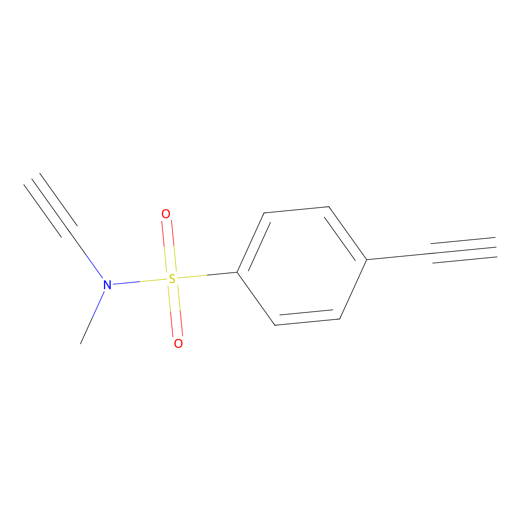 |
100.00 | LDD0444 | [3] | |
|
STPyne Probe Info |
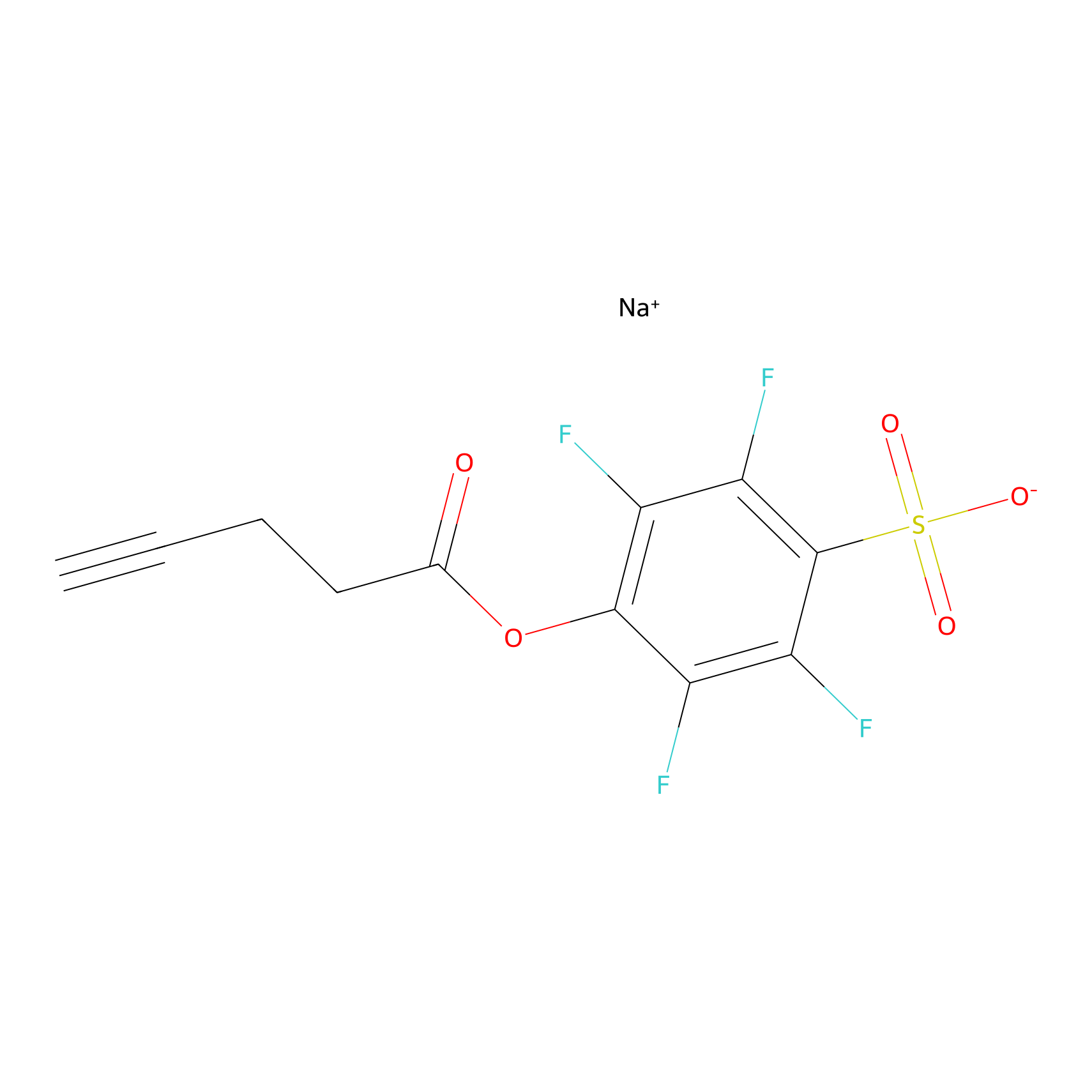 |
K101(6.67); K165(3.51) | LDD0277 | [4] | |
|
ONAyne Probe Info |
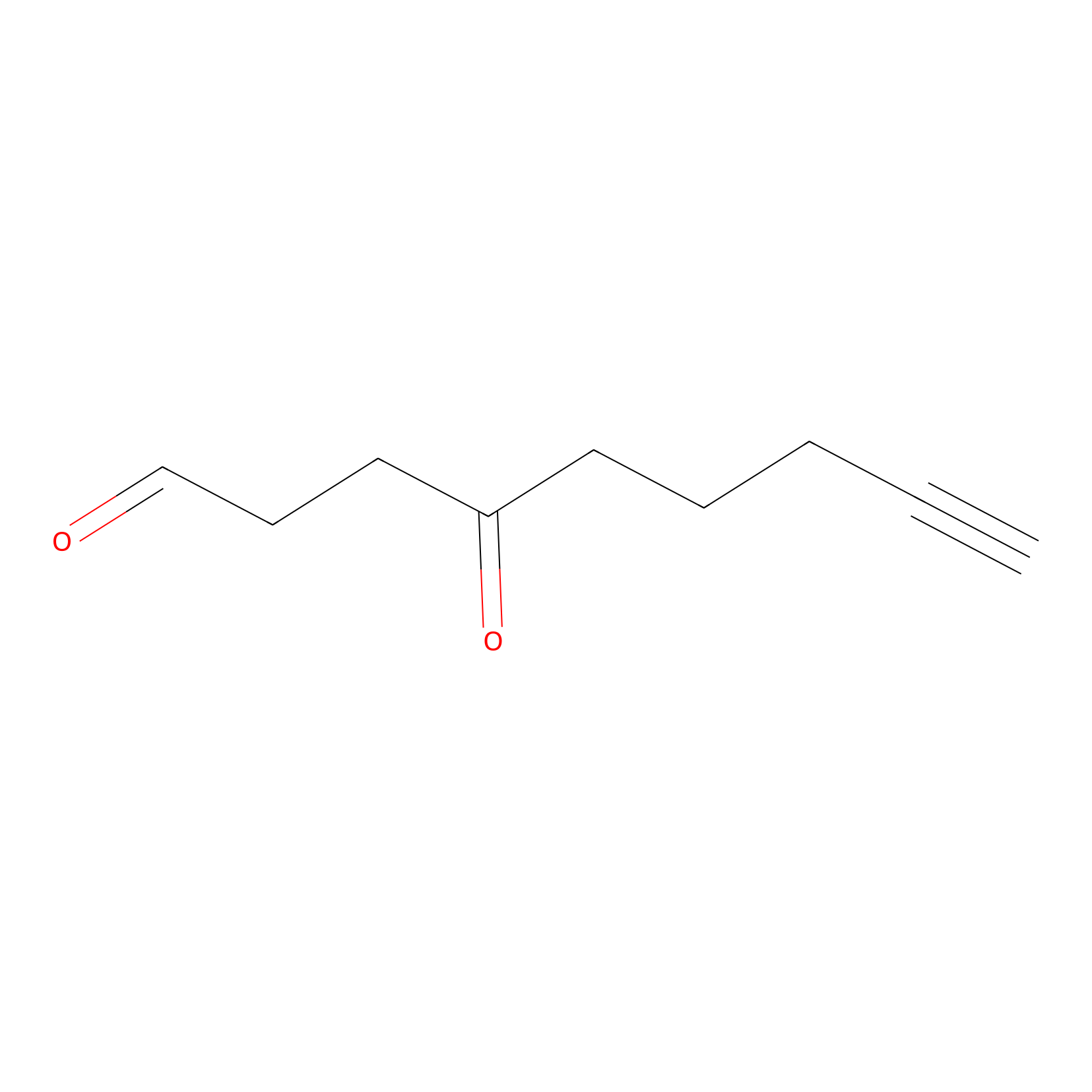 |
K182(0.00); K165(0.00); K143(0.00) | LDD0273 | [4] | |
|
IPM Probe Info |
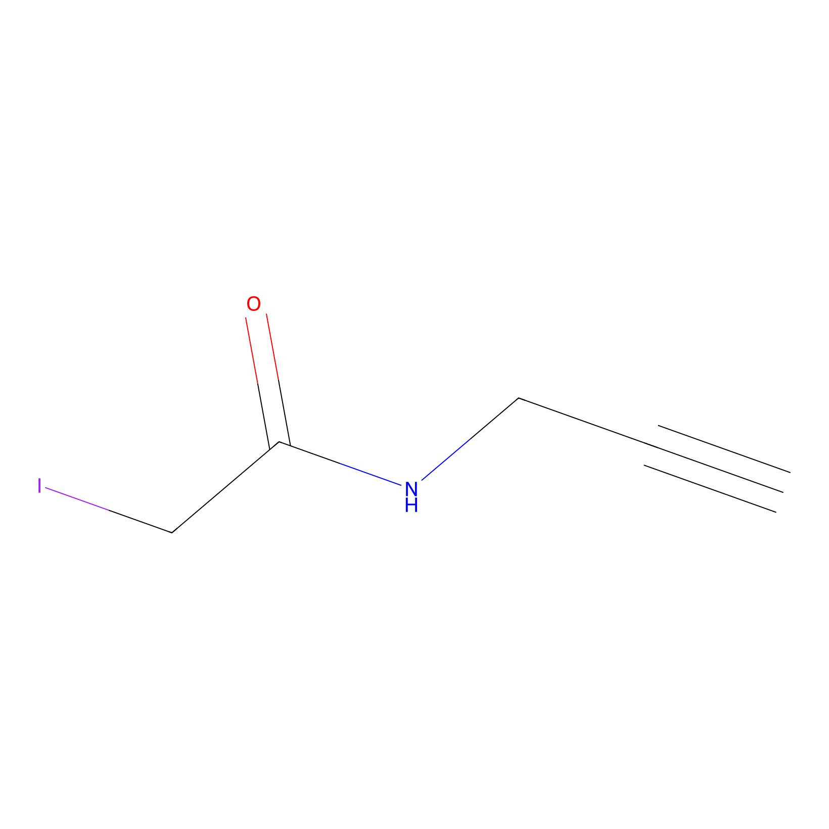 |
N.A. | LDD0241 | [5] | |
|
Probe 1 Probe Info |
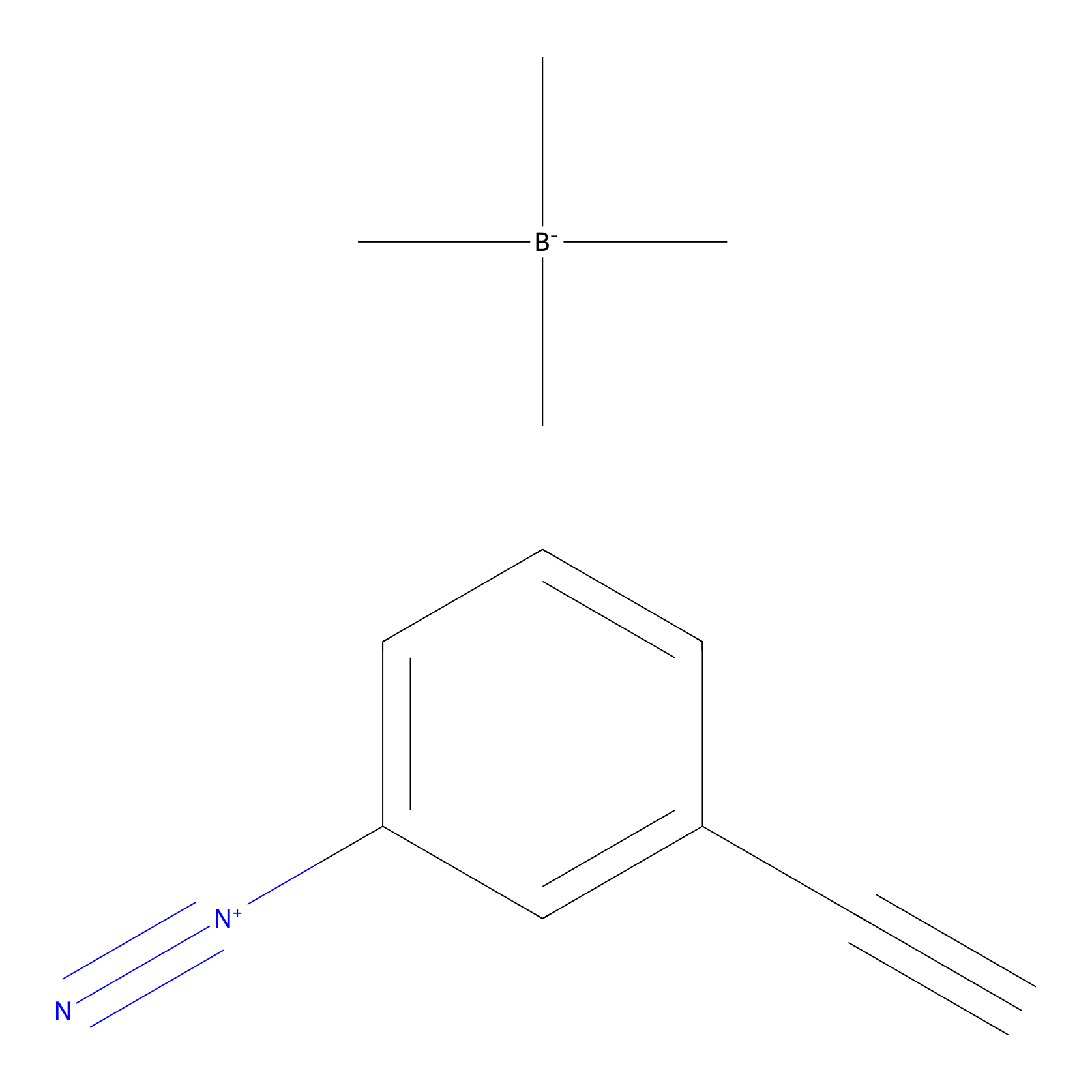 |
Y82(9.75) | LDD3495 | [6] | |
|
DBIA Probe Info |
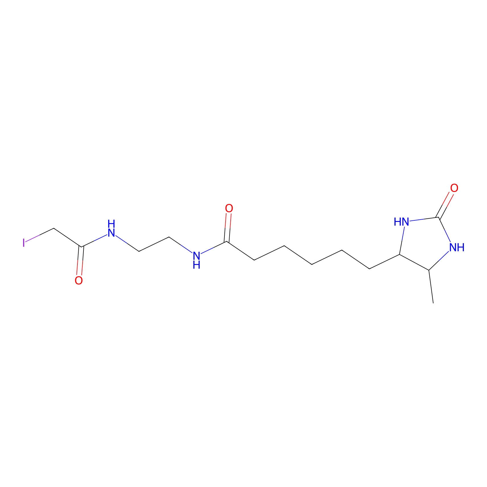 |
C121(1.13) | LDD3313 | [7] | |
|
BTD Probe Info |
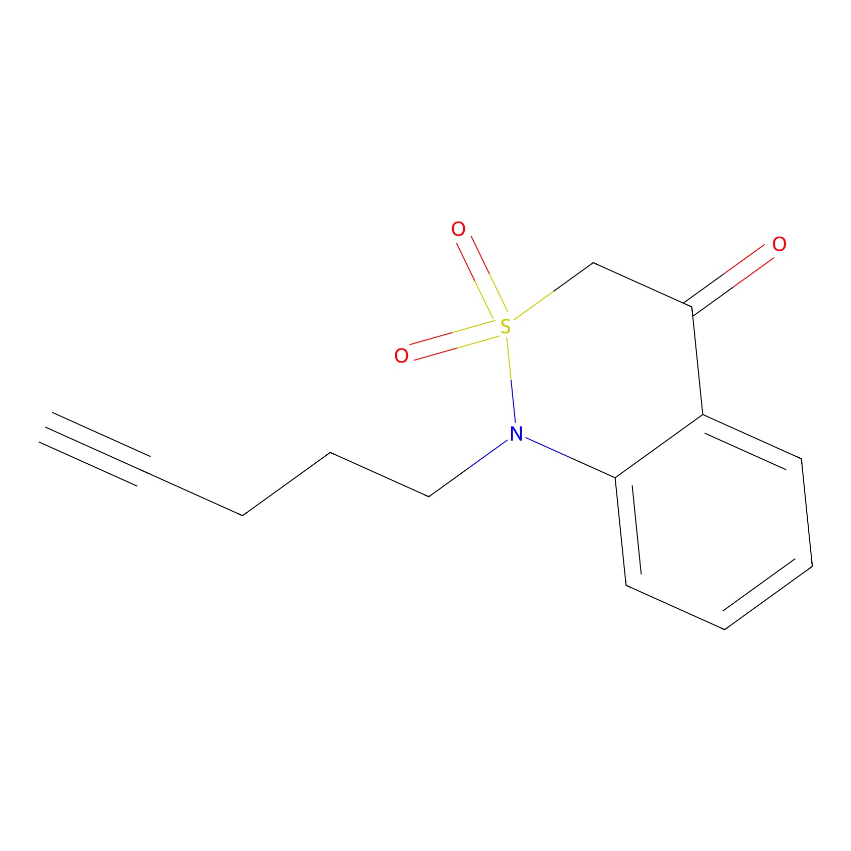 |
C61(0.85) | LDD2089 | [8] | |
|
ATP probe Probe Info |
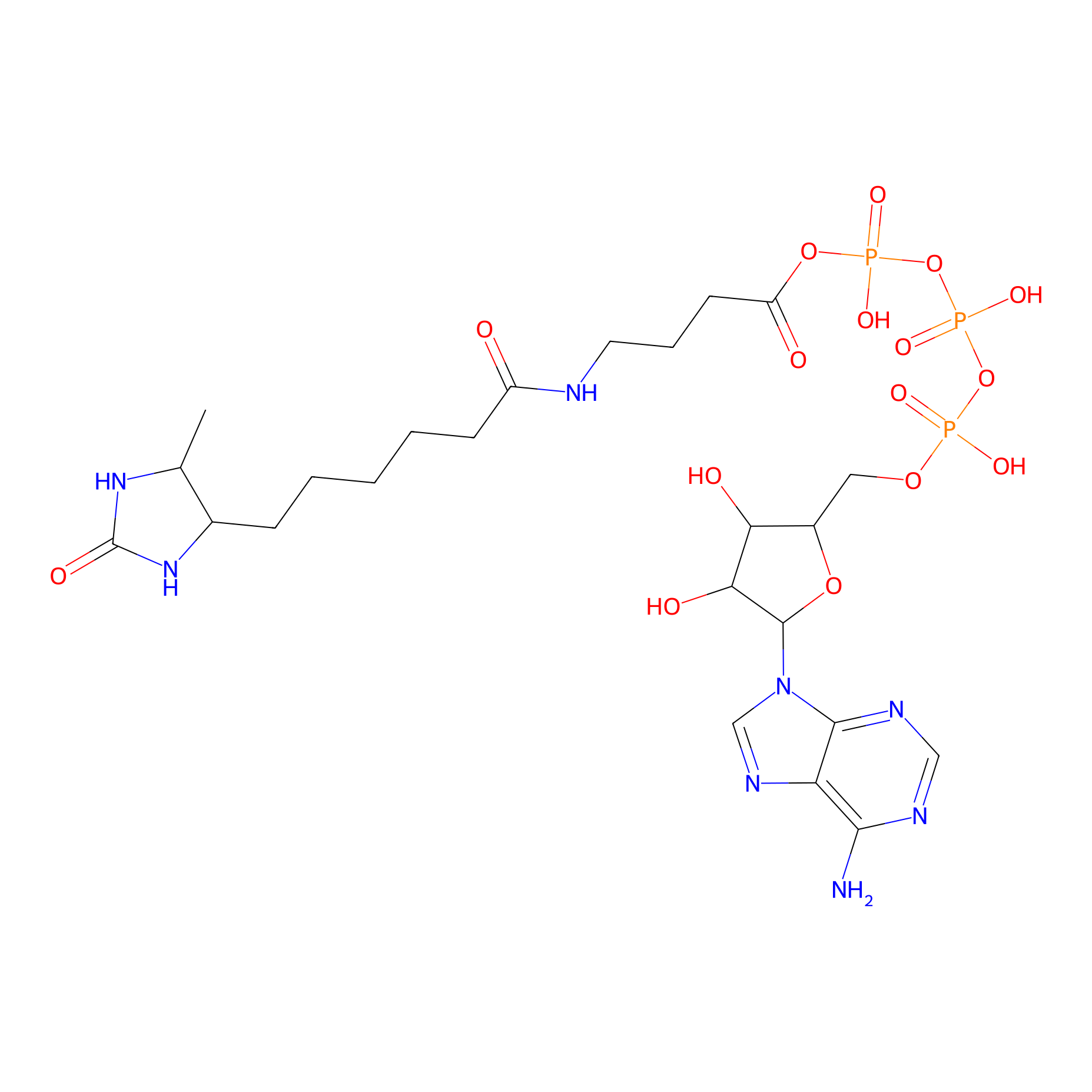 |
K58(0.00); K165(0.00) | LDD0199 | [9] | |
|
m-APA Probe Info |
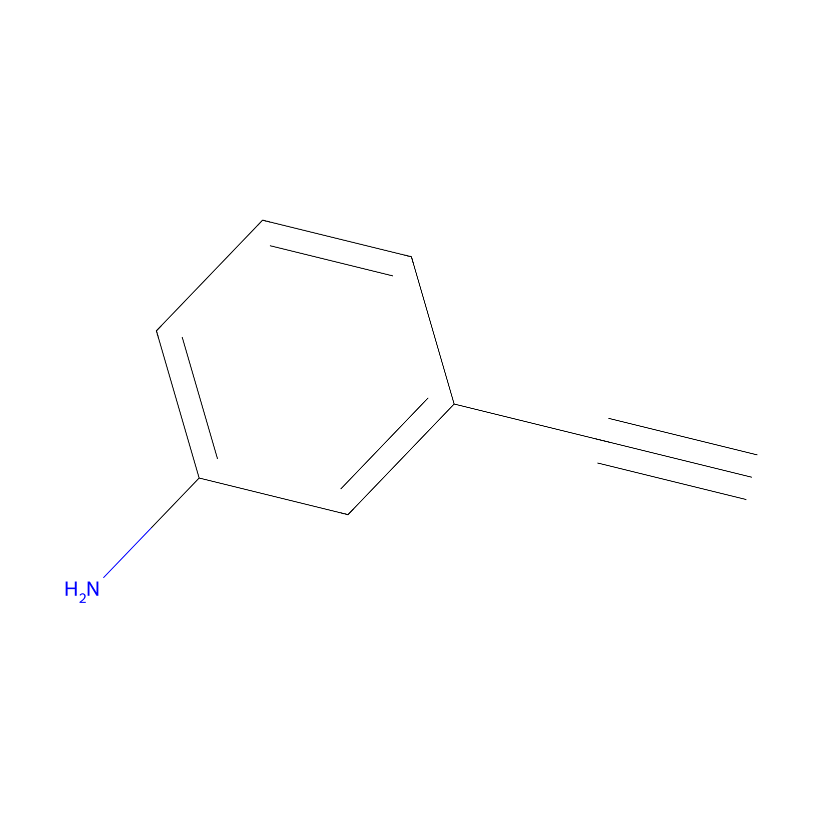 |
N.A. | LDD2231 | [10] | |
|
4-Iodoacetamidophenylacetylene Probe Info |
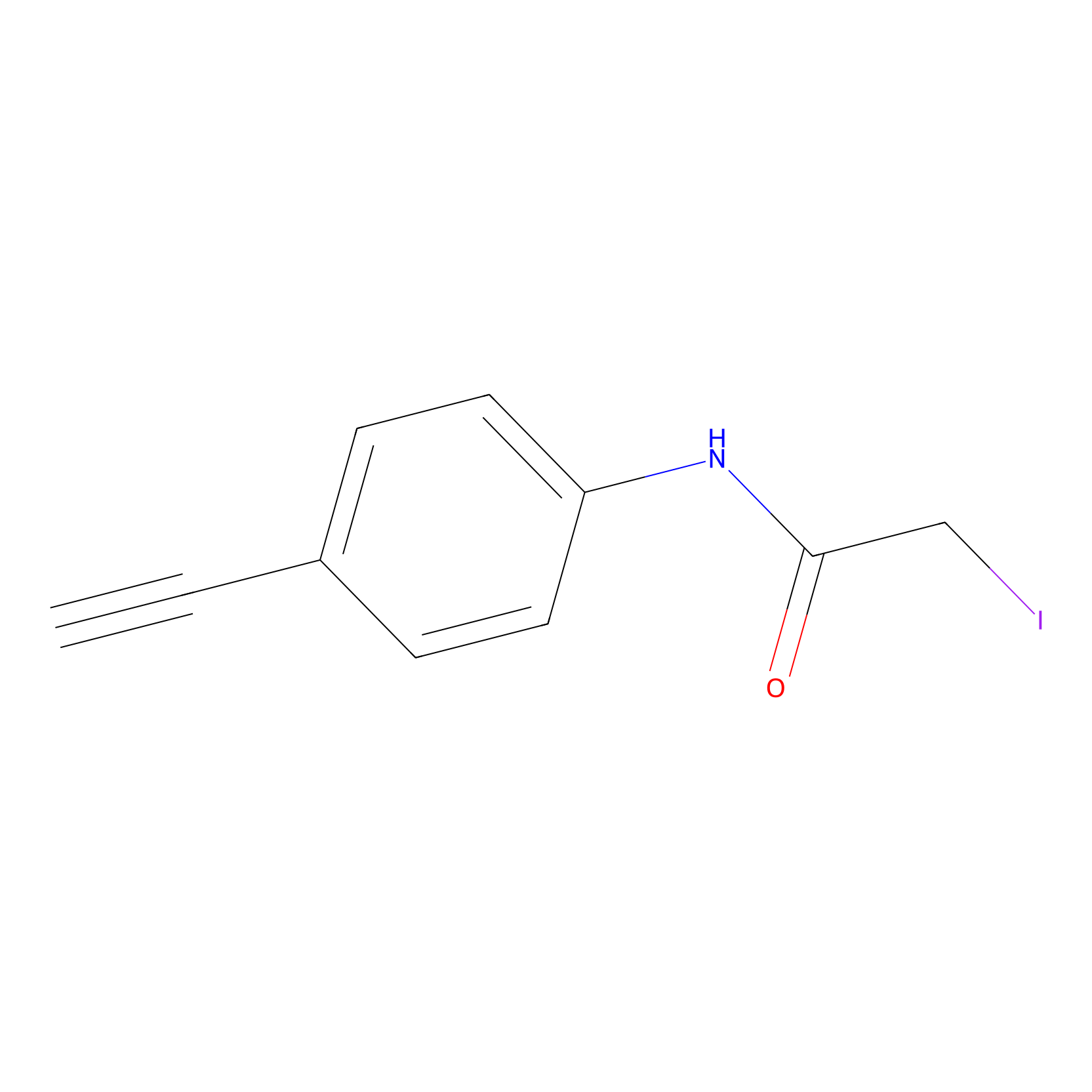 |
N.A. | LDD0038 | [11] | |
|
IA-alkyne Probe Info |
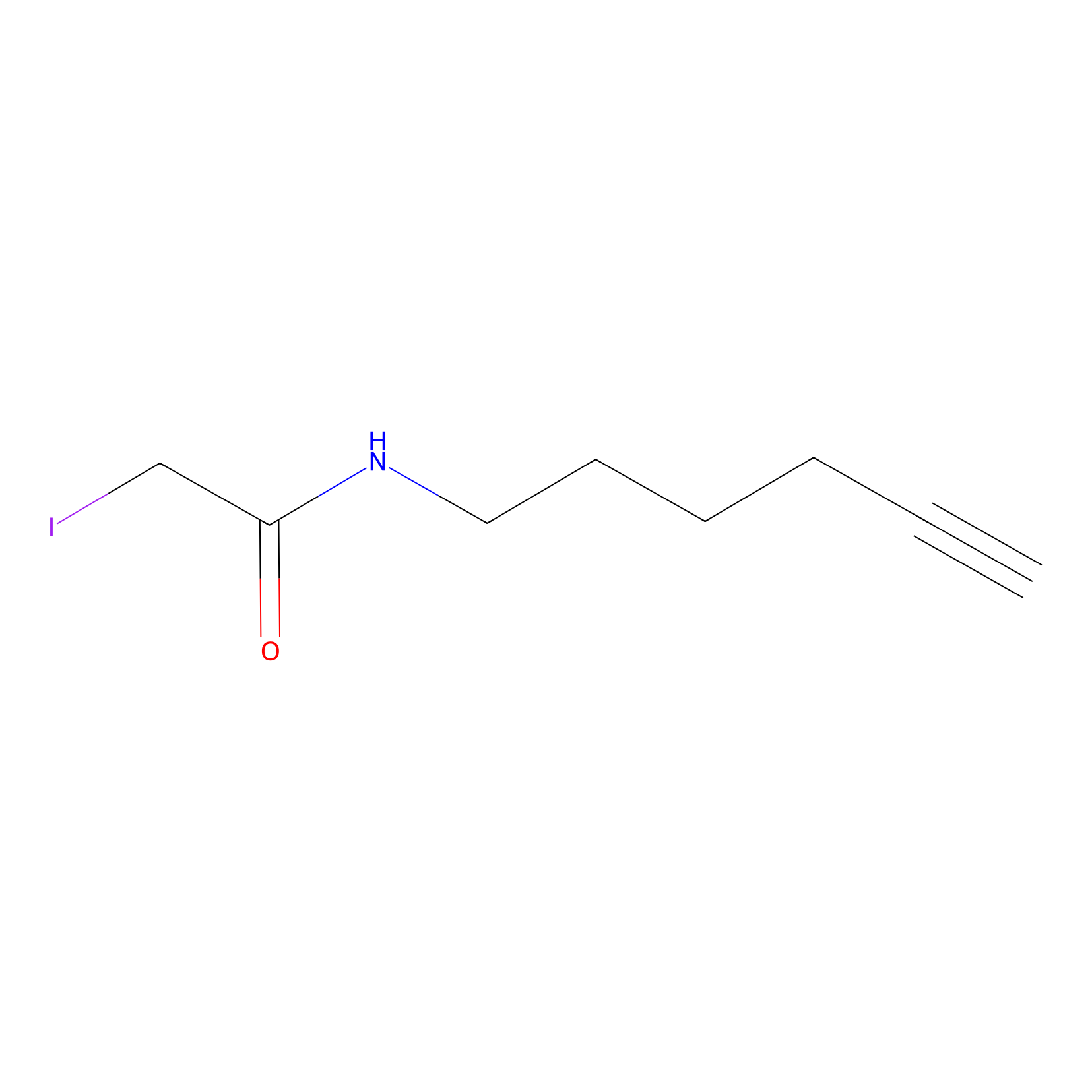 |
N.A. | LDD0036 | [11] | |
|
IPIAA_H Probe Info |
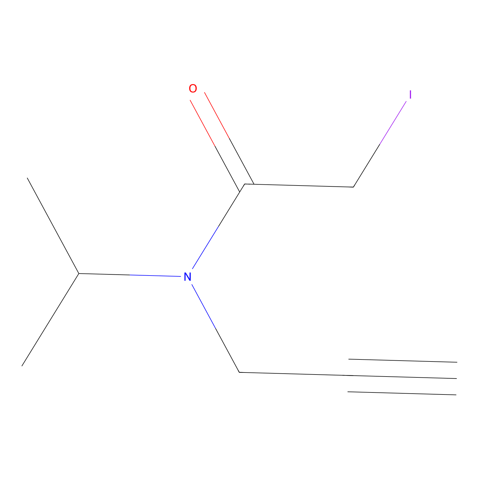 |
N.A. | LDD0030 | [12] | |
|
IPIAA_L Probe Info |
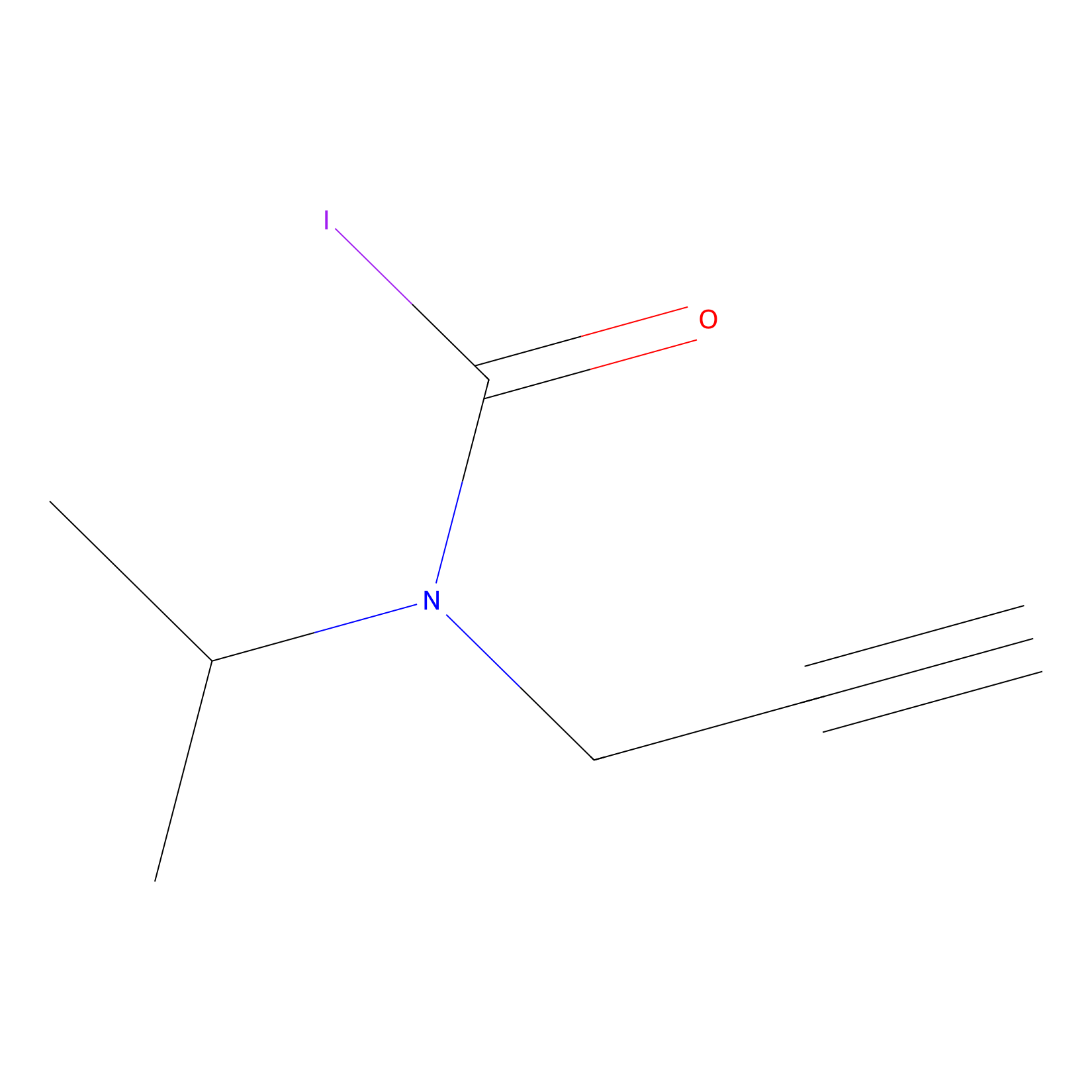 |
C121(0.00); C61(0.00) | LDD0031 | [12] | |
|
ATP probe Probe Info |
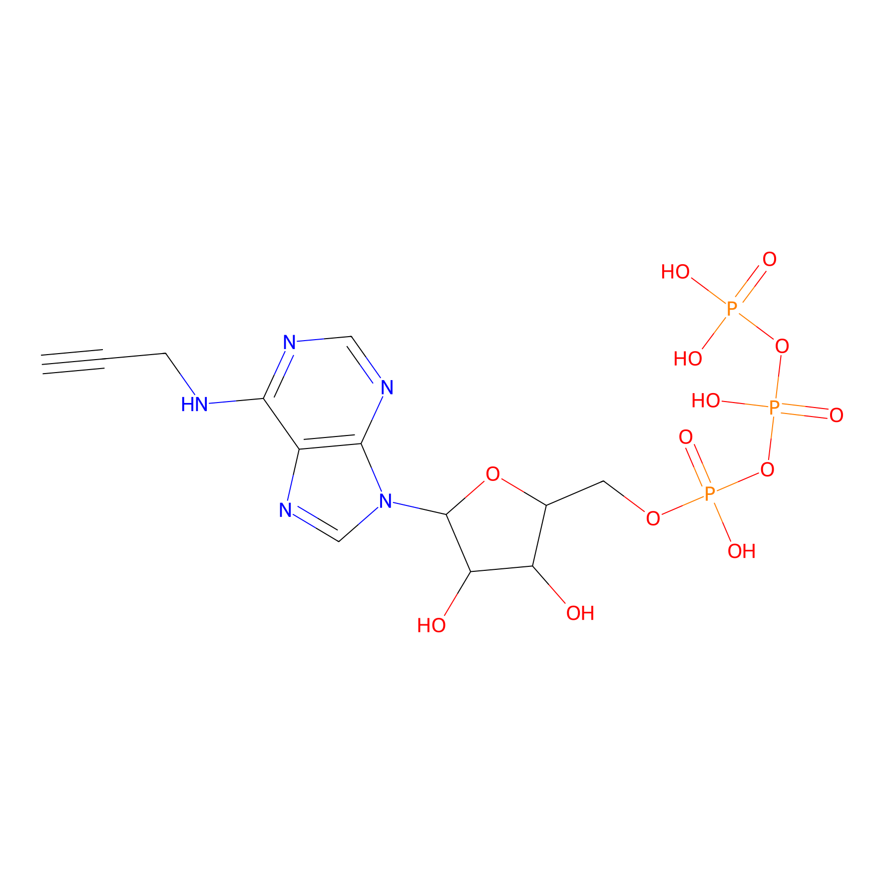 |
N.A. | LDD0035 | [13] | |
|
NAIA_4 Probe Info |
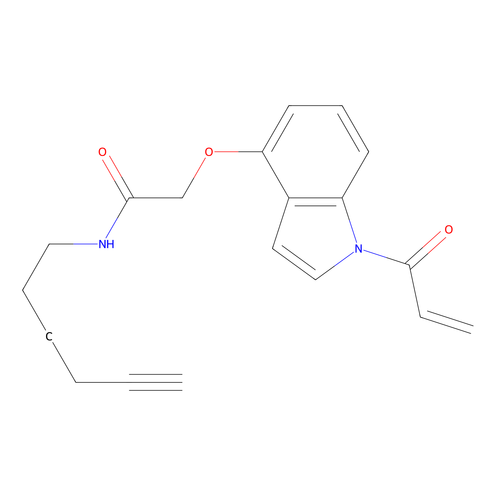 |
N.A. | LDD2226 | [14] | |
|
NPM Probe Info |
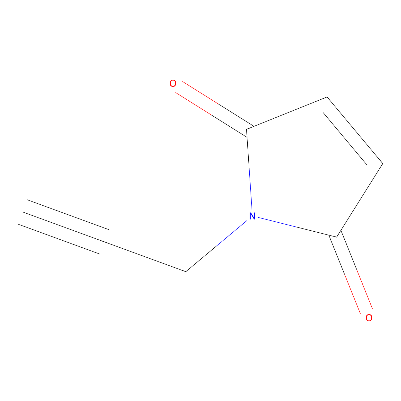 |
N.A. | LDD0016 | [15] | |
|
SF Probe Info |
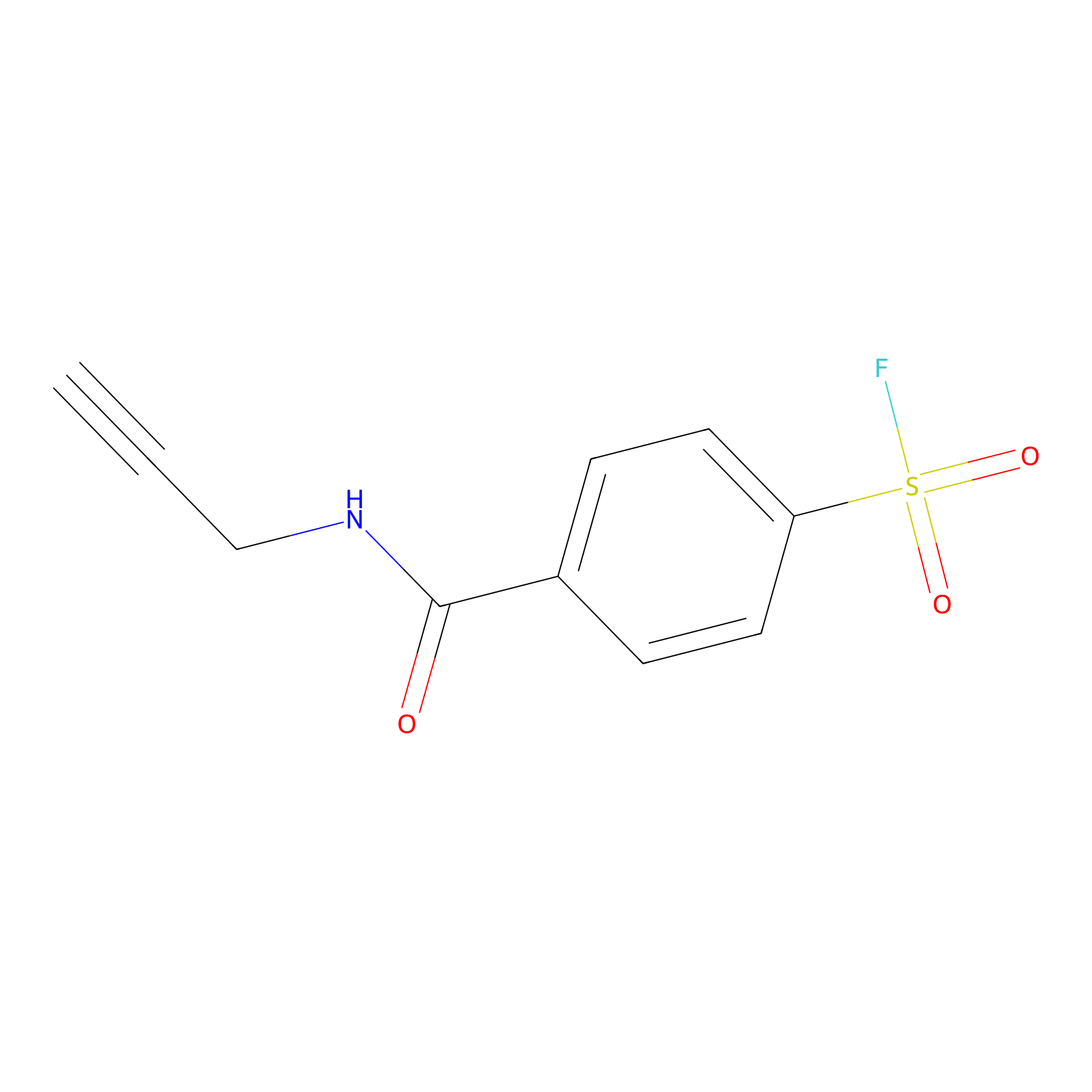 |
Y91(0.00); Y269(0.00); Y53(0.00); Y102(0.00) | LDD0028 | [16] | |
|
TFBX Probe Info |
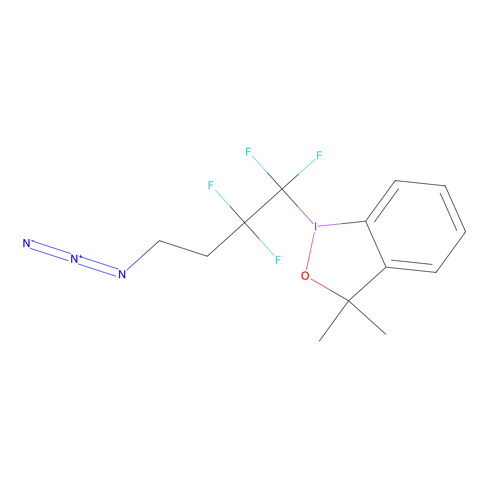 |
C61(0.00); C121(0.00) | LDD0148 | [17] | |
|
1c-yne Probe Info |
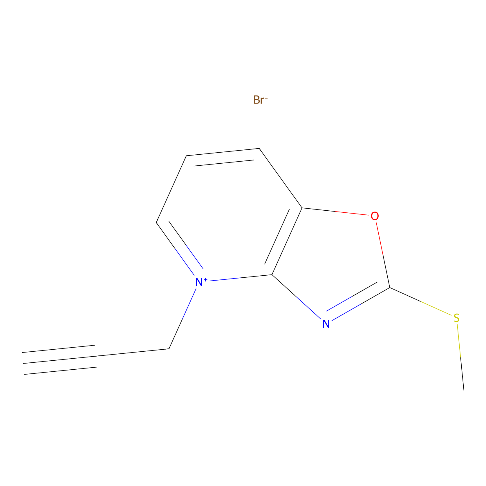 |
N.A. | LDD0228 | [18] | |
|
Acrolein Probe Info |
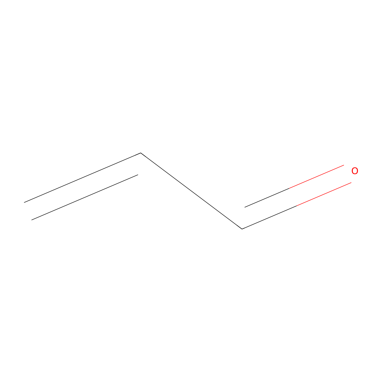 |
H69(0.00); C61(0.00) | LDD0217 | [19] | |
|
Crotonaldehyde Probe Info |
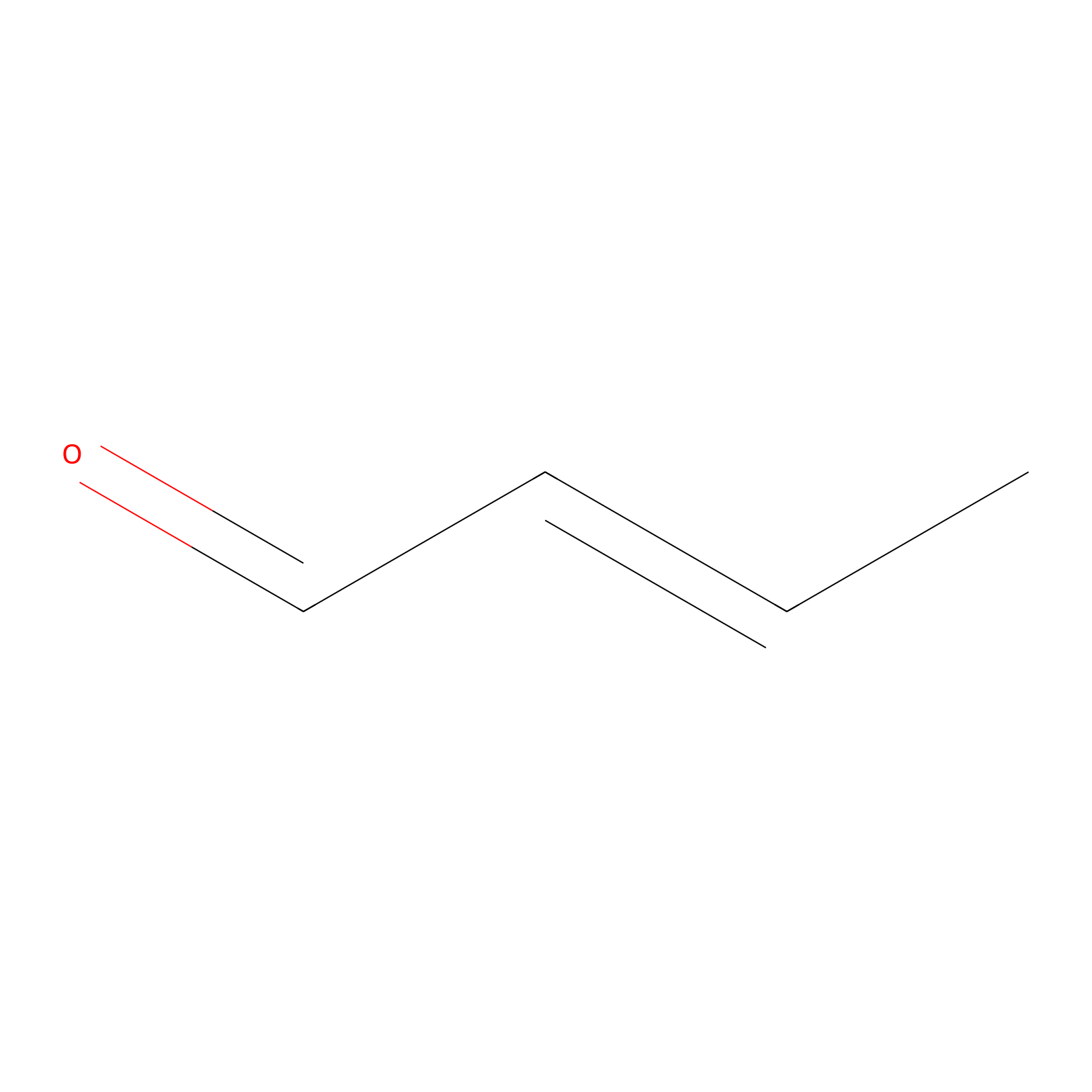 |
C121(0.00); C61(0.00) | LDD0219 | [19] | |
|
Methacrolein Probe Info |
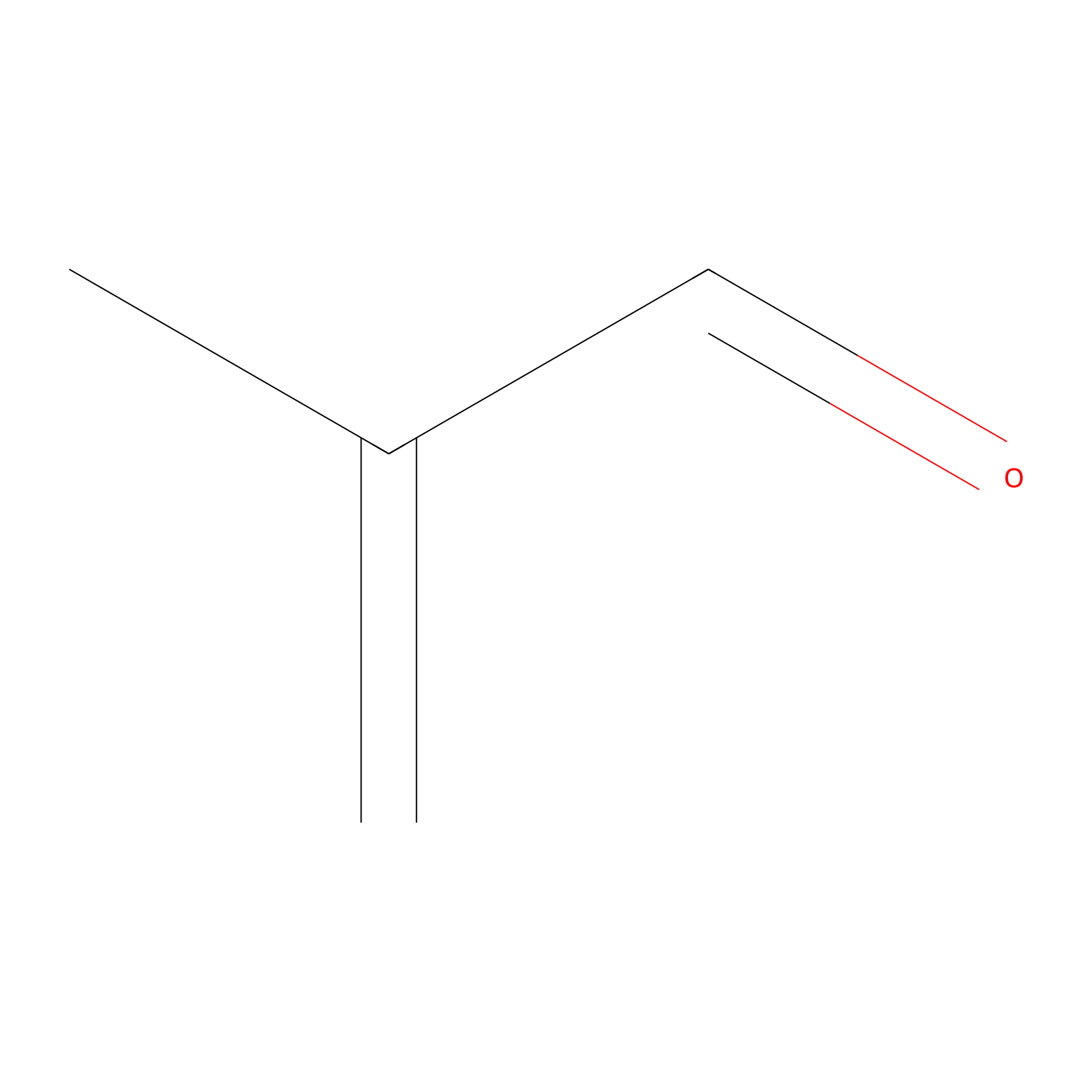 |
N.A. | LDD0218 | [19] | |
|
NAIA_5 Probe Info |
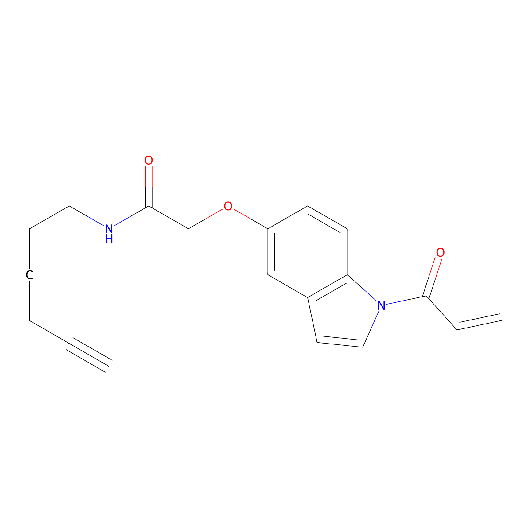 |
C121(0.00); C61(0.00) | LDD2223 | [14] | |
|
HHS-482 Probe Info |
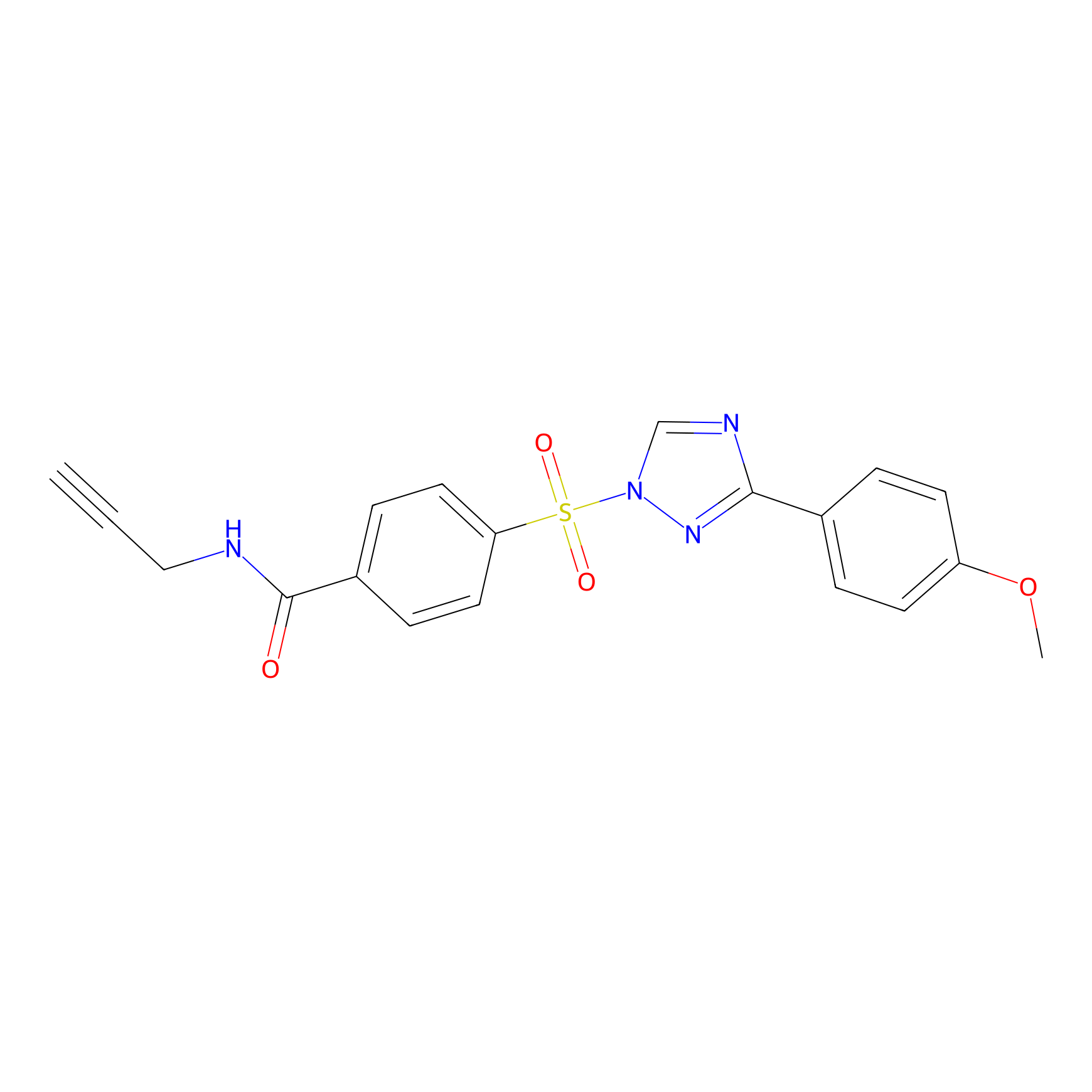 |
Y37(0.85) | LDD2239 | [20] | |
PAL-AfBPP Probe
| Probe name | Structure | Binding Site(Ratio) | Interaction ID | Ref | |
|---|---|---|---|---|---|
|
STS-1 Probe Info |
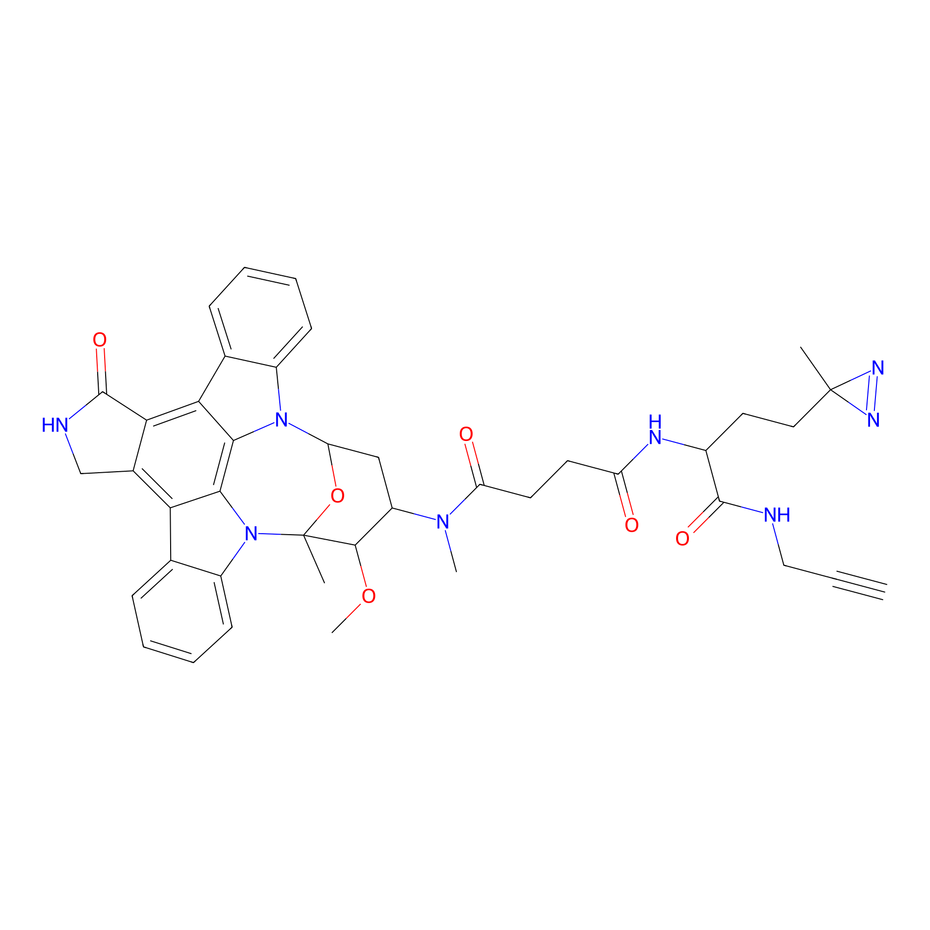 |
N.A. | LDD0137 | [21] | |
|
STS-2 Probe Info |
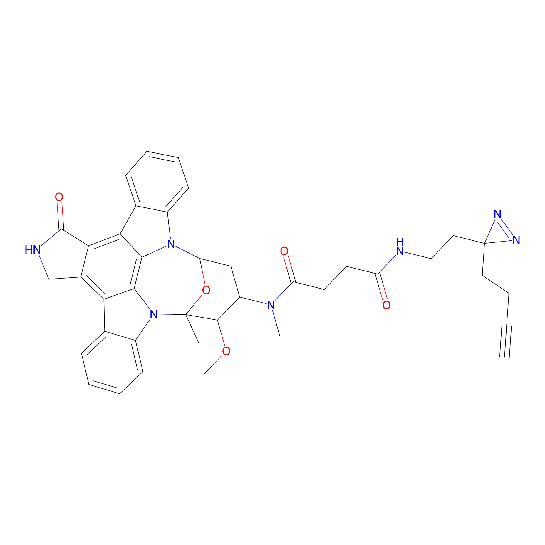 |
N.A. | LDD0138 | [21] | |
|
Staurosporine capture compound Probe Info |
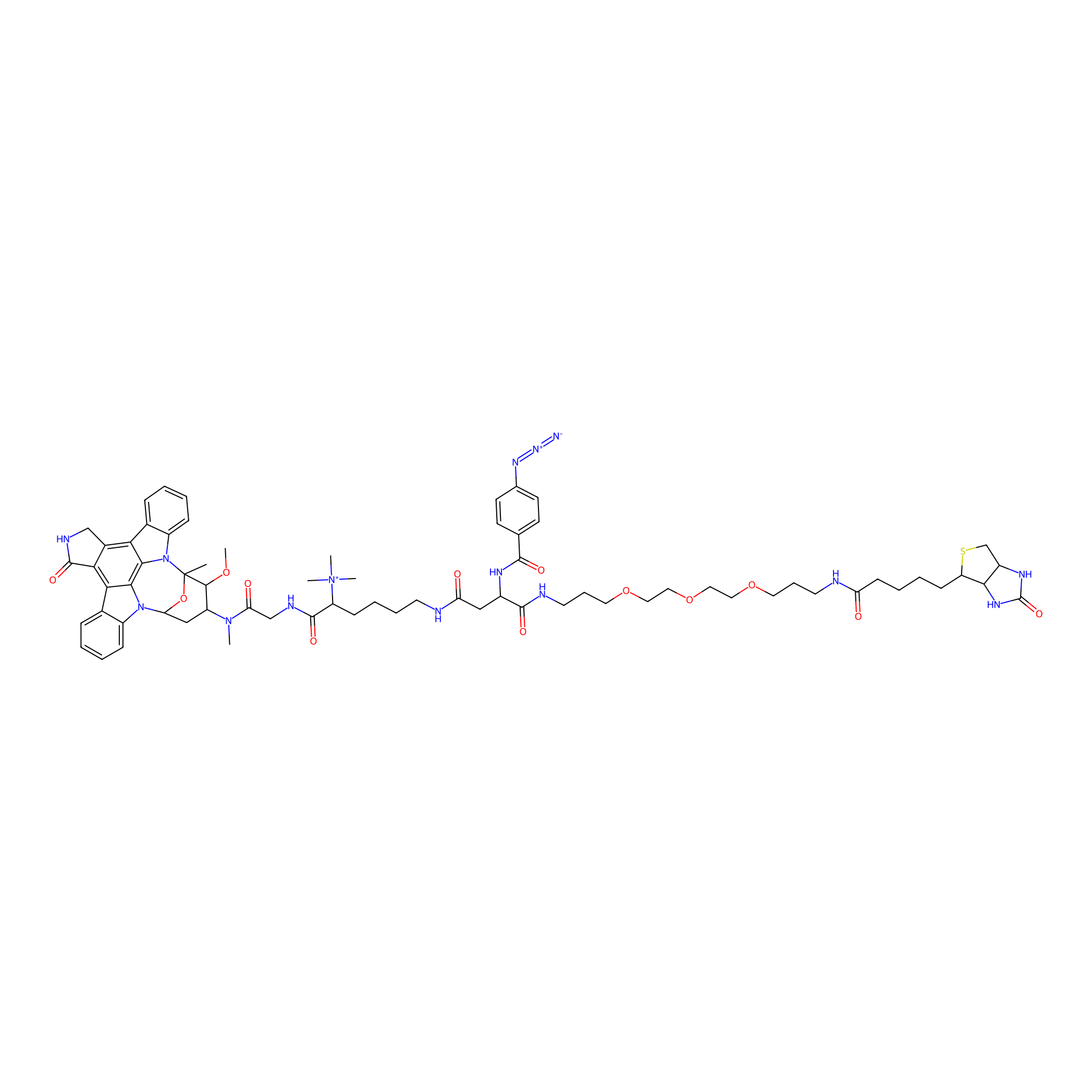 |
N.A. | LDD0083 | [22] | |
Competitor(s) Related to This Target
| Competitor ID | Name | Cell line | Binding Site(Ratio) | Interaction ID | Ref |
|---|---|---|---|---|---|
| LDCM0548 | 1-(4-(Benzo[d][1,3]dioxol-5-ylmethyl)piperazin-1-yl)-2-nitroethan-1-one | MDA-MB-231 | C61(0.53) | LDD2142 | [8] |
| LDCM0519 | 1-(6-methoxy-3,4-dihydroquinolin-1(2H)-yl)-2-nitroethan-1-one | MDA-MB-231 | C61(1.19) | LDD2112 | [8] |
| LDCM0502 | 1-(Cyanoacetyl)piperidine | MDA-MB-231 | C61(0.93) | LDD2095 | [8] |
| LDCM0537 | 2-Cyano-N,N-dimethylacetamide | MDA-MB-231 | C121(0.99) | LDD2130 | [8] |
| LDCM0524 | 2-Cyano-N-(2-morpholin-4-yl-ethyl)-acetamide | MDA-MB-231 | C61(1.19); C121(1.10) | LDD2117 | [8] |
| LDCM0510 | 3-(4-(Hydroxydiphenylmethyl)piperidin-1-yl)-3-oxopropanenitrile | MDA-MB-231 | C61(0.84) | LDD2103 | [8] |
| LDCM0108 | Chloroacetamide | HeLa | H69(0.00); C121(0.00); C61(0.00) | LDD0222 | [19] |
| LDCM0213 | Electrophilic fragment 2 | MDA-MB-231 | C121(0.98) | LDD1702 | [8] |
| LDCM0625 | F8 | Ramos | C121(0.62) | LDD2187 | [23] |
| LDCM0572 | Fragment10 | Ramos | C121(0.77) | LDD2189 | [23] |
| LDCM0573 | Fragment11 | Ramos | C121(0.11) | LDD2190 | [23] |
| LDCM0574 | Fragment12 | Ramos | C121(0.66) | LDD2191 | [23] |
| LDCM0575 | Fragment13 | Ramos | C121(0.58) | LDD2192 | [23] |
| LDCM0576 | Fragment14 | Ramos | C121(0.74) | LDD2193 | [23] |
| LDCM0579 | Fragment20 | Ramos | C121(0.83) | LDD2194 | [23] |
| LDCM0580 | Fragment21 | Ramos | C121(0.71) | LDD2195 | [23] |
| LDCM0582 | Fragment23 | Ramos | C121(1.31) | LDD2196 | [23] |
| LDCM0578 | Fragment27 | Ramos | C121(1.37) | LDD2197 | [23] |
| LDCM0586 | Fragment28 | Ramos | C121(1.15) | LDD2198 | [23] |
| LDCM0588 | Fragment30 | Ramos | C121(1.39) | LDD2199 | [23] |
| LDCM0589 | Fragment31 | Ramos | C121(1.36) | LDD2200 | [23] |
| LDCM0590 | Fragment32 | Ramos | C121(0.63) | LDD2201 | [23] |
| LDCM0468 | Fragment33 | Ramos | C121(1.14) | LDD2202 | [23] |
| LDCM0596 | Fragment38 | Ramos | C121(0.33) | LDD2203 | [23] |
| LDCM0566 | Fragment4 | Ramos | C121(0.73) | LDD2184 | [23] |
| LDCM0614 | Fragment56 | Ramos | C121(0.65) | LDD2205 | [23] |
| LDCM0569 | Fragment7 | Ramos | C121(0.68) | LDD2186 | [23] |
| LDCM0571 | Fragment9 | Ramos | C121(0.75) | LDD2188 | [23] |
| LDCM0107 | IAA | HeLa | H69(0.00); H142(0.00); C61(0.00) | LDD0221 | [19] |
| LDCM0022 | KB02 | Ramos | C121(0.48) | LDD2182 | [23] |
| LDCM0023 | KB03 | MDA-MB-231 | C121(1.92) | LDD1701 | [8] |
| LDCM0024 | KB05 | Hs 936.T | C121(1.13) | LDD3313 | [7] |
| LDCM0109 | NEM | HeLa | H142(0.00); H69(0.00) | LDD0223 | [19] |
| LDCM0496 | Nucleophilic fragment 11a | MDA-MB-231 | C61(0.85) | LDD2089 | [8] |
| LDCM0505 | Nucleophilic fragment 15b | MDA-MB-231 | C61(0.82) | LDD2098 | [8] |
| LDCM0506 | Nucleophilic fragment 16a | MDA-MB-231 | C61(1.34); C121(1.38) | LDD2099 | [8] |
| LDCM0507 | Nucleophilic fragment 16b | MDA-MB-231 | C61(0.59) | LDD2100 | [8] |
| LDCM0511 | Nucleophilic fragment 18b | MDA-MB-231 | C121(0.49) | LDD2104 | [8] |
| LDCM0514 | Nucleophilic fragment 20a | MDA-MB-231 | C61(1.22); C121(1.11) | LDD2107 | [8] |
| LDCM0515 | Nucleophilic fragment 20b | MDA-MB-231 | C61(1.15) | LDD2108 | [8] |
| LDCM0521 | Nucleophilic fragment 23b | MDA-MB-231 | C121(0.36) | LDD2114 | [8] |
| LDCM0527 | Nucleophilic fragment 26b | MDA-MB-231 | C61(0.79) | LDD2120 | [8] |
| LDCM0530 | Nucleophilic fragment 28a | MDA-MB-231 | C61(1.02); C121(1.01) | LDD2123 | [8] |
| LDCM0531 | Nucleophilic fragment 28b | MDA-MB-231 | C61(0.45) | LDD2124 | [8] |
| LDCM0532 | Nucleophilic fragment 29a | MDA-MB-231 | C61(1.04); C121(0.84) | LDD2125 | [8] |
| LDCM0533 | Nucleophilic fragment 29b | MDA-MB-231 | C61(0.80) | LDD2126 | [8] |
| LDCM0535 | Nucleophilic fragment 30b | MDA-MB-231 | C61(0.86) | LDD2128 | [8] |
| LDCM0543 | Nucleophilic fragment 38 | MDA-MB-231 | C121(1.25) | LDD2136 | [8] |
| LDCM0544 | Nucleophilic fragment 39 | MDA-MB-231 | C61(0.99) | LDD2137 | [8] |
| LDCM0546 | Nucleophilic fragment 40 | MDA-MB-231 | C61(0.98) | LDD2140 | [8] |
| LDCM0549 | Nucleophilic fragment 43 | MDA-MB-231 | C61(0.96) | LDD2143 | [8] |
| LDCM0550 | Nucleophilic fragment 5a | MDA-MB-231 | C61(3.70) | LDD2144 | [8] |
| LDCM0551 | Nucleophilic fragment 5b | MDA-MB-231 | C61(1.54) | LDD2145 | [8] |
| LDCM0019 | Staurosporine | Hep-G2 | N.A. | LDD0083 | [22] |
The Interaction Atlas With This Target
References
